A Crash Course on Shoulder Anatomy

Introduction

Diagnosis of shoulder pathologies is a difficult and specialised field in physiotherapy. As such, instead of diving straight into some shoulder pathologies that can be overlooked and misdiagnosed, I have decided to take a step back and discuss the important anatomical considerations. These points are common knowledge to your physiotherapist- but should serve the active sportsperson and weekend warrior due reminder to take care of a complex and easily damaged structure.

Glenohumeral Stability

Ball Seal

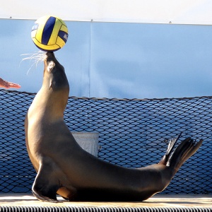
The Glenohumeral (GH) joint is designed for the extremes of mobility, which unfortunately comes at the sacrifice of the stability that many other joints- such as the hip joint- take for granted. Compared with the deep ball and socket joint of the hip, the shoulder joint better reflects a sea lion balancing a ball on its nose (Brukner & Kahn 2012)! This analogy stems from the extremely small and shallow glenoid fossa in relation to the oversized head of the humerus.

The unstable nature of this joint highlights the huge importance of static and dynamic restraints in the shoulder for both stability and function. Now, static restraints refer to those which permit stability of the shoulder but do not aid in movement. These include the joint capsule, which is made slightly deeper by a circumferential lip called the glenoid labrum and the various GH ligaments. Conversely, dynamic stabilisers have dual functions in assisting with stability and movement of the shoulder joint. Dynamic stabilisers refer to the muscles of the rotator cuff- the supraspinatus, infraspinatus, teres minor and subscapularis muscles. These muscles arise on the scapula- each have different actions (see table 1.1) and engulf the shoulder with their insertions onto the head of the humerus. They then function to co-contract simultaneously, thereby maintaining the humeral head in the glenoid fossa (Brukner & Kahn 2012).

Muscle Movement
Supraspinatus Aids abduction in the first 30-degrees
Infraspinatus Chief external rotator of the arm
Teres Minor External rotation- aids in extension
Subscapularis Internal rotation

*Note: These muscles- principally the supraspinatus muscle work in unison to prevent the humeral head from translating superiorly when the arm is raised.

Scapulohumeral Rhythm

Normal movement of the shoulder requires fluent action at four different joints:

  • Scapulothoracic Joint: Movement of the scapula, gliding on the rib cage. The attachment of this joint is purely muscular. Movements occurring here include elevation/depression, retraction/protraction and superior/inferior rotation.
  • Acromioclavicular Joint: Movement about this joint is very slight- but this synovial joint actually allows small amounts of superior and inferior glide.
  • Sternoclavicular Joint: This refers to the joint of the acromion at the manubrium of the sternum. Movements allowed here include elevation/depression, anterior/posterior translation and small amounts of rotation.
  • Glenohumeral Joint: Movement of the head of the humerus in the glenoid fossa.

Full movement of the shoulder requires some movement occurring at all of these joints. Collectively, this movement is termed scapulohumeral rhythm. Impaired movement at any of these joints will manifest itself as poorly controlled, jerky movement of the shoulder.

Disturbances to Scapulohumeral Rhythm

Scapular Muscles

The common causes and manifestations of disturbed scapulohumeral rhythm will be discussed in further damage as I examine common pathologies of the shoulder joint. That said- the most common cause is weakness and poor motor control of the scapula stabilising muscles. The stable and optimally placed scapula ensures that the muscles arising from the scapula (i.e. the rotator cuff muscles) maintain their adequate length and thereby tension as they act on the head of the humerus. This highlights the importance of training not only the dynamic restraints of the shoulder, but also the scapula stabilising muscles following any shoulder injury.

References:

Brukner, P & Kahn, K 2012, ‘Brukner & Kahn’s Clinical Sports Medicine’, 4th edn, McGraw Hill, Sydney.
Illiotibial Band Friction Syndrome

Introduction

Illiotibial Band Friction Syndrome (ITBFS) is the most common cause of lateral knee pain. It is commonly experienced by athletes who participate in sports with continual repetitive motion of the knee- especially runners and cyclists. The athlete will present with tenderness on the lateral knee, inferior to the lateral epicondyle of the femur and superior to the joint line.

The most common differential diagnosis for this condition is tendinopathy of the Biceps Femoris. This is generally predicated by excessive downhill running or running on an uneven surface and most commonly experienced by football and soccer players who will also experience pain with kicking and sprinting. If lateral knee pain is experienced following an acute ankle injury, it may indicate the superior tibiofibular joint or lateral meniscus as the site of the injury.

This article will focus on ITBFS. It will discuss the relevant anatomy of the knee joint, the pathology of the condition and management options.

ITB

Anatomy

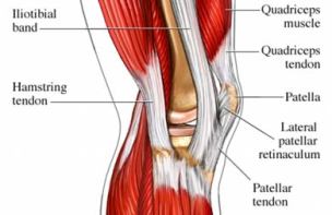
  • The ITB should not be considered a muscle. Instead, it is a lateral thickening of the fascia which envelopes the whole thigh. It receives muscular insertions from the Tensor Fascia Latae muscle and also from the Gluteus Maximus muscle.
  • The ITB has a number of insertions. It has connections with the Linea Aspera along the lateral surface of the femur. It has distal attachments to the lateral femoral epicondyle, lateral surface of the patella and the lateral epicondyle of the Tibia.
  • The ITB functions predominantly for stability at the knee and hip joint. At the knee joint- the ITB reinforces the lateral collateral ligament of the knee.
  • Underlying the ITB at the lateral epicondyle of the Femur is a richly innervated layer of fat and connective tissue. This is the site of irritation in ITBFS.
  • The Biceps Femoris (hamstring) passes posteriorly to the lateral epicondyle of the femur and inserts into the head of the Fibula. Palpation of this structure is important when ruling out the differential diagnosis of Biceps Femoris tendinopathy.

Pathology of the Condition

It was previously believed that ITBFS resulted from friction as occurs through anterior and posterior movement of the ITB in knee flexion and extension respectively. However recent research now identifies that strong fibrous bands of connective tissue around the distal portion of the ITB prevents this movement. Instead, it is now suggested that altering tension of the Tensor Fascia Latae and other hip musculature results in compressive forces around the lateral femoral epicondyle. Pain is therefore thought to result from repetitive compressive loads to the fat pad overlying the lateral epicondyle.

Contributing Factors

Generally speaking, ITBFS is predicated by poor sporting technique and biomechanical abnormalities. The biomechanical factors that can contribute to this pathology include:

  1. Weak Hip Abductors: This results in greater hip adduction during single-leg stance in walking and running. As a result, there is greater strain on the ITB and the structures underneath (Lavine 2010).
  2. Tight ITB: This is the most logical causative factor- as a tighter ITB means greater compression of the tissues underneath. Ober’s test often reveals tightness of the ITB and can produce a burning sensation. Further investigation is then required to determine whether this tightness results from proximal tightness of the TFL or Gluteus Maximus muscles, or distal over-development of the Vastus Lateralis (Brukner et. al 2012).
  3. Angle of Knee Flexion in Stance Phase: The compression generated by the ITB on lateral structures distally is greatest when the knee if flexed between 20-30 degrees. It is for this reason that running downhill and on uneven surfaces can severely exacerbate ITBFS. These activities require a greater amount of knee flexion during locomotion (Lavine 2010).

Treatment of ITB Friction Syndrome

Treatment should address not only the local pathology at the lateral knee, but also foot and hip biomechanics that could be contributing to greater stress on the ITB.

  1. In the case of acute exacerbation- ice, electrotherapy modalities and non-steroidal anti-inflammatory drugs may provide pain relief and limit inflammation.
  2. Soft-tissue treatment, such as dry needling, myofacial release and self-massage with the foam roller are all used to release tension in the ITB.
  3. Stretches of the ITB are regularly prescribed- but because the ITB has attachments along the length of the femur at the Linea Aspera these stretches are mostly ineffective. However, stretching of the TFL and of the Gluteus Maximus muscle can reduce tension in the ITB due to their insertions into this sheath.
  4. To continue on from the above point- trigger point therapy of the ITB and gluteal muscles can release tension in the ITB.
  5. Strengthening exercises of the hip abductors should also be prescribed in the presence of weakness. This can reduce the amount of hip adduction which may occur in single-leg stance and thereby reduce pressure on the lateral epicondyle by the ITB.
  6. If conservative management fails- surgical release may be indicated.

References:

Lavine, R 2010, ‘Illiotibial band friction syndrome’, Current reviews in musculoskeletal medicine, vol. 3, no. 1, pp. 18-25.

Brukner, P & Kahn, K 2012, ‘Brukner & Kahn’s Clinical Sports Medicine’, 4th edn, McGraw Hill, Sydney.